
Artistry and creativity don't have to be and shouldn't be separate from science. There is so much beauty in the natural world and science allows us to see patterns and symmetry in structures at all levels: from microscopic to light years away.
The relationship between form and function is something that has always fascinated me. To see an image of a neuron compared to: a section of a cerebellum, a tree, and a view of a river from space (see images below). They all have a symmetrical branching pattern and one could almost be mistaken for another under the right circumstance. Dendrite, a term of Greek origin that refers to a tree, is the scientific name for neuron branches. Dendritic drainage is used to describe the pattern of branching of a body of water. Scientist seek out these similar forms and then try to find a relation between their functions. They can then make predictions about where this pattern may be found again or what the function is of something that follows this pattern.
There is beauty and wonder in this world and science is a great way to explore it.
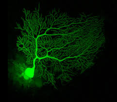
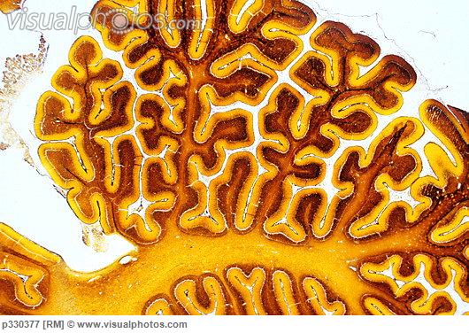
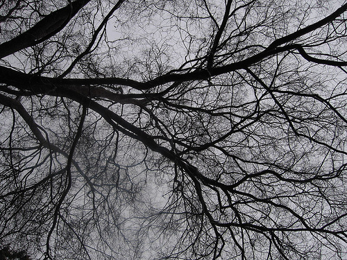
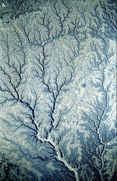
above left: rat Purkinje neuron; above right: human cerebellum; bottom left: elm tree; bottom right: rivers and streams in the republic of South Yemen
Taken from:
http://www.rikenresearch.riken.jp/eng/hom/6236
http://www.visualphotos.com/image/1x6009515/sectioned_brain_cerebellum_light_micrograph
http://www.photoree.com/photos/permalink/819948-56707737@N00
http://cmex.ihmc.us/CMEX/data/catalog/OutflowChannels/Drainage.html
Taken from:
http://www.rikenresearch.riken.jp/eng/hom/6236
http://www.visualphotos.com/image/1x6009515/sectioned_brain_cerebellum_light_micrograph
http://www.photoree.com/photos/permalink/819948-56707737@N00
http://cmex.ihmc.us/CMEX/data/catalog/OutflowChannels/Drainage.html
Scanning Electron Microscope (SEM)

Zacharias and Hans Jansen are accredited for inventing the microscope ~1590. Anton van Leeuwenhoek (1632-1723) and Robert Hooke (1635-1703) each modified the concept and made great contributions to the field of Biology.
New technology has led to vast improvements in microscopy over the years and now scientists have tools such as the scanning electron microscope (SEM). The SEMs allows scientists to see the 3 dimensional shape of an object in fascinating detail.
Advanced microscopes such as the SEM produce such spectacular images that there are even cell imaging competitions! The following are a few of my favorite SEM images.
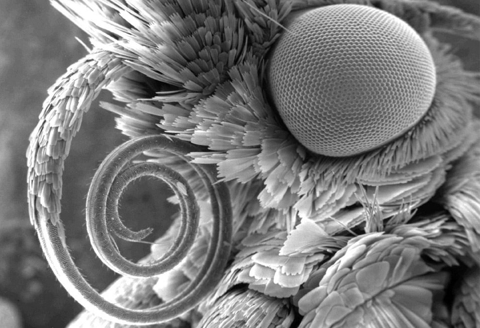
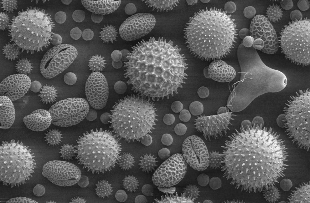
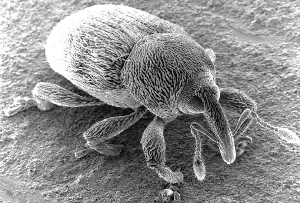
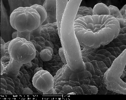
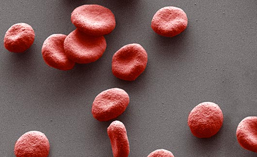
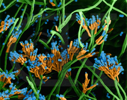
upper left: moth; upper right: pollen; middle left: weevil; middle right: underside of a leaf; bottom left: red blood cells; bottom right: fungi mycelium
Taken from:
http://www.boston.com/bigpicture/2008/11/peering_into_the_micro_world.html
http://remf.dartmouth.edu/Juglans_nigra_walnut_SEM_P2/
Taken from:
http://www.boston.com/bigpicture/2008/11/peering_into_the_micro_world.html
http://remf.dartmouth.edu/Juglans_nigra_walnut_SEM_P2/
Florescence Microscopy

Florescence Microscopy allows scientist to target certain features of cells with fluorescent tags so that when viewed by special microscopes such as the confocal laser scanning microscopes (CLSM), the images glow with beautiful colors and patterns.
The primary difference between CLSM imaging and SEM imaging is that with the CLSM, you are viewing in interior of the specimen and not the surface as in SEM. Another difference is that the color you see in SEM images is always added after by computers whereas most or all of the colour in fluorescence microscopy is due to the special staining and tagging process.
Below are a few of my favorite florescence microscopy images.
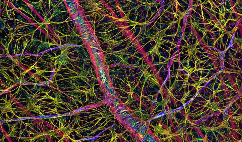
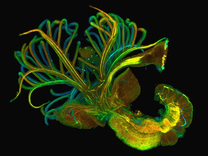

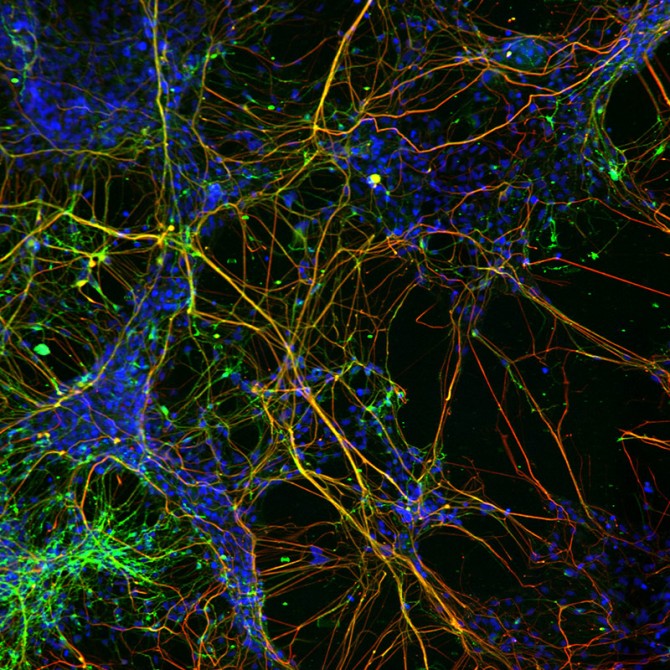
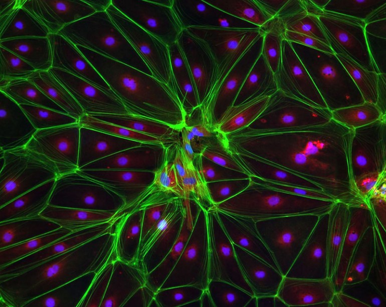
upper left: fruit fly larvae; upper right: filamentous green algae; middle left: aquatic worm; middle right: dividing cancer cell; bottom left: Trisomy 21 neural cells; bottom right: Huntington's disease stem cells
Taken from:
http://www.gizmag.com/cell-imaging-competition-stunning-images/26460/pictures#10
http://ramanan50.wordpress.com/tag/fluorescence-microscope/
http://www.perkinelmer.com/pages/020/cellularimaging/default.xhtml
http://discovermagazine.com/galleries/zen-photo/n/nikon2006-2#.UeBMEhYTHzJ
Taken from:
http://www.gizmag.com/cell-imaging-competition-stunning-images/26460/pictures#10
http://ramanan50.wordpress.com/tag/fluorescence-microscope/
http://www.perkinelmer.com/pages/020/cellularimaging/default.xhtml
http://discovermagazine.com/galleries/zen-photo/n/nikon2006-2#.UeBMEhYTHzJ
